

Data visualization
graphs and scientific figures
Working with researchers at Campbell Laboratory in the University of Iowa I design figures to accompany their work when submitted for publishing. The goal of these images is to provide professional looking images that are also easy to interpret.

Carver College of Medicine
Kevin P. Campbell
Investigator, Howard Hughes Medical Institute
Chair Department of Molecular Physiology & Biophysics
Roy J. and Lucille A. Carver Biomedical Research Chair
Professor, Neurology
Director, Senator Paul D. Wellstone MDSRC

SOON TO BE PUBLISHED:
AMEYA WALIMBE


POMK regulates dystroglycan function via LARGE1-mediated elongation of matriglycan
Walimbe AS, Okuma H, Joseph a, Yang T, Yonekawa T, Hord JM , Venzke D, Anderson ME, Torelli S, Manzur A, Devereaux M, Cuellar M, Prouty S, Ocampo Landa S, Yu L, Xiao J, Dixon JE, Muntoni F, Campbell KP
Synthesis of the α-DG Laminin-Binding Modification and Enzymes Involved.
Synthesis of the laminin-binding modification begins with the addition of the core M3 trisaccharide (GalNAc-β3-GlcNAc-β4-Man) on α-DG by the sequential actions of Protein O-Mannosyltransferase 1 and 2 (POMT1/2), Protein O-linked Mannose N-Acetyl-glucosaminyltransferase 2 (POMGNT2), and β1,3-N-Acetylgalactosaminyltransferase 2 (B3GALNT2), in the ER. POMK phosphorylates the C6 hydroxyl of mannose after synthesis of core M3. The phosphorylated core M3 is further elongated in the Golgi by Fukutin (FKTN), Fukutin related protein (FKRP), Transmembrane Protein 5 (TMEM5), β1,4-Glucuronyltransferase 1 (B4GAT1), and Like-acetyl-glucosaminyltranserase 1 (LARGE1). Isoprenoid synthase domain-containing (ISPD) produces cytidine diphosphate (CDP)-ribitol in the cytosol, and this serves as a sugar donor for the reactions catalyzed by FKTN and FKRP. LARGE1 synthesizes matriglycan, which directly interacts with the LG domains of matrix ligands.
Before:
Dr. Walimbe provided past examples of similar figures, but was looking for an updated and modern approach for their paper. General structure and typical iconography used within the scientific community was utilized.


These figures were created by several different scientists over the span of several years. Many images were old screenshots with the original file lost or difficult to retrieve. Most were created in PowerPoint and they lacked cohesion as a group. The new figures were expected to meet a high standard of professional quality.
After:
Dr. Walimbe explained that many times these images will be seen on a screen and that being able to read the text clearly on multiple devices was important. Large 18pt text was used to increase legibility and accessibility.
Texture (based on the microscopic imaging) was referenced to bring emphasis and detail to the composition. Color was used to break apart the image and bring focus to the different areas and make clear which process were the most closely related.


Dystroglycanopathy research is the study of specific muscles and the mutations that can occur within individuals. These studies are used to help develop therapeutic strategies for patients with muscular dystrophy. It was important that I was able to understand some of the mechanisms and interactions taking place in these figures, so that I could accurately depict what was required.
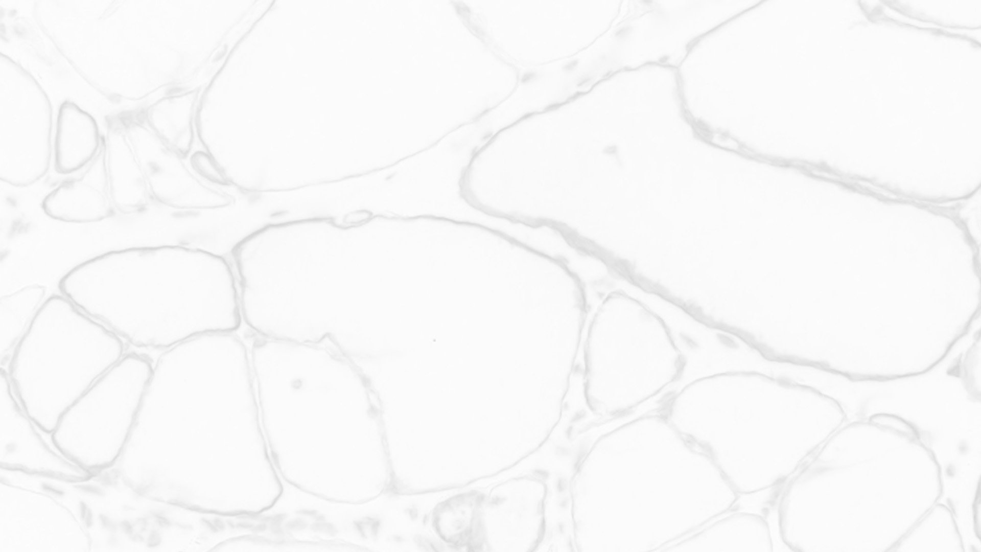
As a visual storyteller I found microscopic images to be enlightening and they were used as a reference for the textures. The texture of the muscle and the synaptic background help to tell a story about the scale of these figures. Using 3-dimensional illustrations also helped establish a hierarchy within the figure’s narrative.


Consistent iconography and an easy to read key helped tie these figures together.

This project pushed me to familiarize myself with ways to create small files with large amounts of detail.
A combination of Adobe Photoshop and Illustrator were used. While I previously had experience using Adobe’s shape builder tool, some of these images required further research into best practices.
PUBLISHED:

HIDEHIKO OKUMA
N-terminal domain on dystroglycan enables LARGE1 to extend matriglycan on α-dystroglycan and prevents muscular dystrophy
Okuma H, Hord JM, Chandel I, Venzke D, Anderson ME, Walimbe AS, Joseph S, Gastel Z, Hara Y, Saito F, Matsumura K, Campbell KP
Domain structure of dystroglycan (DG) and Δ-α-DGN.
Wild-type DG is a pre-proprotein with an N-terminal signal peptide (light green) that is translated in the rough endoplasmic reticulum. The globular N-terminal domain (α-DGN; orange) is present in wild-type DG but absent in the mutant (∆-α-DGN). The junction between α-DGN and the mucin-like domain (light teal) contains a furin convertase site. The globular extracellular C-terminal domain (CTD; pink) contains an SEA (sea urchin sperm protein, enterokinase and agrin) autoproteolysis site, which cleaves pro-DG into α-DG and β-DG (green). Glycosylation has been omitted for clarity.
Before:
A collection of images, sketches created in PowerPoint and data points were provided to convey the general idea of what the figures would include.


Raw images of western blots and data points were used as a starting point for many of the images.
After:
Images were cropped and edited so that data points were easier to read. Sketches were converted into 3D structures and a layout was developed to help create clarity. Most of this project included creating simple, clean and easy to read graphs while adhering to the dimensional requirements for each figure.


Explore Rachel’s work:
-
![]()
User Experience
-
![]()
Published images
-
![]()
Clothing & Apparel Design
-
![]()
Photography
-
![]()
Illustration